Stiffness and elasticity: our self-sensing cantilevers can be used to measure the Young’s modulus via nanoindentation and force-distance curves.
Adhesion: pull-off forces between the tip of the self-sensing cantilever and the sample surface can be quantified.
Friction and shear: tracks lateral twisting of the cantilever to measure local friction (Lateral Force Microscopy).
Viscoelasticity: with our self-sensing cantilever, material creep, relaxation, and loss/storage moduli can be evaluated.
In practice
How researchers use it
Example 1: Nanoindentation of graphene, watched live in the SEM
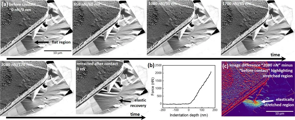
(a) SEM image sequence during AFM force-distance nanoindentation on a suspended few-layer graphene membrane. (b) The corresponding force-distance curve. (c) Colour-coded difference image showing the elastically stretched region of the membrane. Credit: Hummel et al., Appl. Phys. Lett. 118, 103104 (2021), CC BY 4.0
The study. How much does an AFM tip deform an atomically thin membrane while measuring it? Researchers at the University of Vienna and GETec Microscopy answered this by indenting a suspended few-layer graphene membrane and imaging it with the SEM at the same time. The SEM shows the elastically stretched area around the contact.
Where our cantilever is used. The self-sensing cantilever of the AFSEM is both the indenter and the force sensor: it presses on the membrane and records the force-distance curve, while the electron beam watches from above. This only works because the cantilever needs no laser inside the SEM.
S. Hummel et al., Applied Physics Letters 118, 103104 (2021), doi:10.1063/5.0040522. CC BY 4.0.
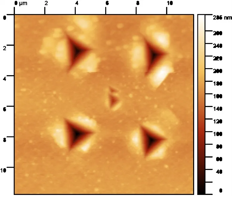
AFM topography of an array of nanoindents in electroplated copper, with a small additional indent placed in the centre. Credit: c-sense and Alemnis
The study. In the Nanomechanical Microscope, developed with Alemnis, a nanoindenter and an AFM share one stage. Indents are placed with sub-micrometre precision and imaged right afterwards. The image shows an array of indents in electroplated copper with a small additional indent placed in the centre.
Where our cantilever is used. A self-sensing cantilever is the AFM part of the instrument. It images each indent directly after it is made; its small, laser-free head is what allows AFM and indenter to sit side by side.
c-sense and Alemnis measurement.
Example 3: Force-distance curves on a single nanowire
SEM view of the cantilever tip on a free-standing nanowire (left) with the measured amplitude-distance and force-distance curves (right). Credit: GETec Microscopy and c-sense
The study. The mechanical response of one individual free-standing nanowire is measured inside the SEM. The tip is placed on the wire under live SEM view, the wire is bent, and the force-distance and amplitude-distance curves are recorded.
Where our cantilever is used. The self-sensing cantilever of the AFSEM pushes on the nanowire and measures the force; the SEM image provides the bending amplitude. With a conductive cantilever the conductivity of the wire can be measured in the same contact.
GETec Microscopy and c-sense measurement (AFSEM).
Discuss your application
Let's discuss your measurement.
Tell us about your setup and we'll help identify the right sensing solution.