Self-sensing cantilevers can be modified with a functionalized magnetic coating or a magnetic FEBID tip to ensure a well-defined magnetic moment, making them ready to use for MFM applications. The sharp tip and integrated readout of the self-sensing cantilevers enable fine topography resolution and reliable magnetic field acquisition at the desired lift height.
In practice
How researchers use it
Example 1: A compact MFM inside a 12 T magnet
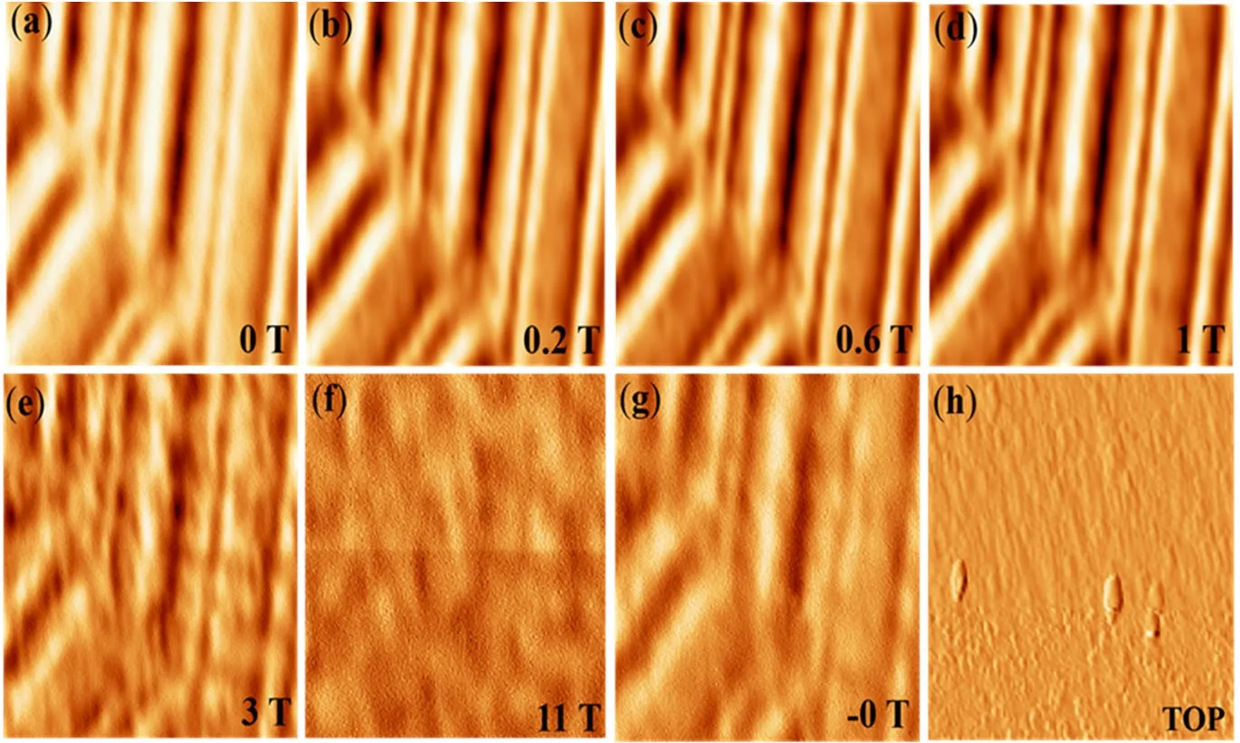
(a to g) Magnetic domains of a videotape at 5 K while the field is raised from 0 to 11 T. (h) Topography. Image size 12 µm × 12 µm. Credit: Abas et al., Micromachines 13, 1922 (2022), CC BY 4.0
The study. The High Magnetic Field Laboratory in Hefei and USTC built a magnetic force microscope that fits into a 12 T cryogen-free superconducting magnet. As a test they imaged the magnetic domains of a videotape at 5 K while raising the field from 0 to 11 T (image size 12 µm × 12 µm).
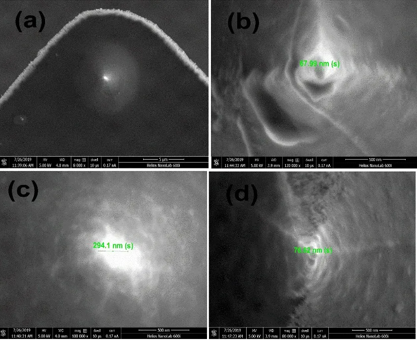
Where our cantilever is used. The force sensor of the microscope is our PRSA-L300-F50-STD cantilever, with a magnetic film coated onto its tip. Because the deflection is read out electrically, the MFM head needs no optics and fits into the narrow magnet bore.
SEM images of the magnetically coated tip of the piezoresistive cantilever at different magnifications. Credit: Abas et al., Micromachines 13, 1922 (2022), CC BY 4.0
A. Abas et al., Micromachines 13, 1922 (2022), doi:10.3390/mi13111922. CC BY 4.0.
Example 2: Magnetic domains written with an AFM tip
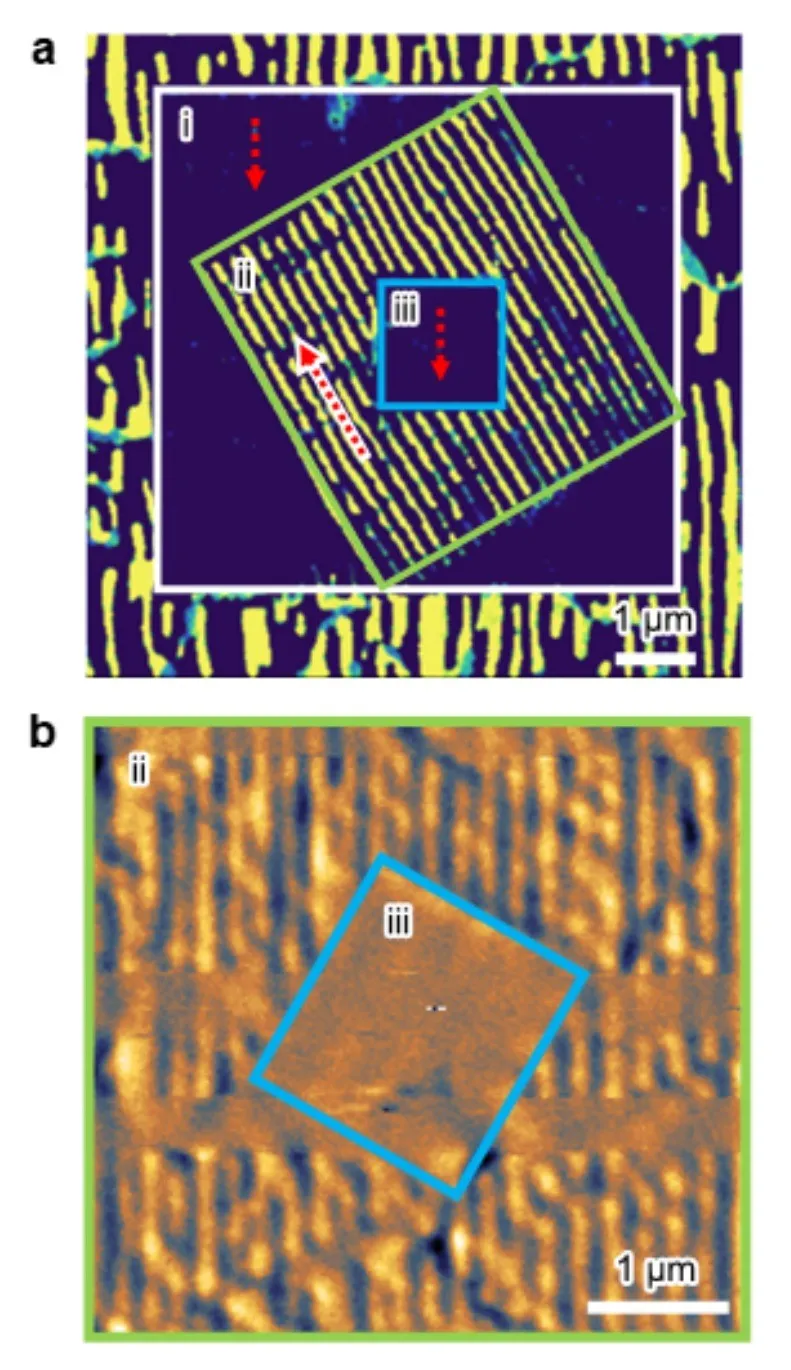
(a) Electron channelling contrast image of crystal domains written into SrRuO₃ with an AFM tip. (b) MFM image of the ferromagnetic domains in the written regions. Scale bars 1 µm. Credit: Peng et al., open preprint of Nature Nanotechnology 20, 1199–1204 (2025), CC BY 4.0
The study. A team from the University of Warwick, the High Magnetic Field Laboratory in Hefei and partners showed that the crystal direction in SrRuO₃ thin films can be written locally with an AFM tip. Since magnetism in this material follows the crystal direction, the written patterns become designed magnetic domains.
Where our cantilever is used. The magnetic domains were imaged by MFM with our PRSA-L300-F50-STD cantilever, coated with Ti, Co and Au to make the tip magnetic. The MFM ran in a 12 T superconducting magnet and, for higher fields, in a 35 T magnet. Panel b shows the MFM image of the written regions.
W. Peng et al., Nature Nanotechnology 20, 1199–1204 (2025), doi:10.1038/s41565-025-01950-z. Figure from the open preprint, CC BY 4.0.